Western Blot Tips
We are dedicated to helping our customers achieve exceptional results. While the information below can never be an exclusive solution to every problem you may encounter; it is our hope that you will find the information useful and even beneficial in troubleshooting any problems you may have. Of course, if you find you are still having problems you may submit them to us using this Western Blot Troubleshooting Form.
SYMPTOMS
- Wrong band size
- No bands/signal
- High uniform background
- Swirly speckled background
- White bands
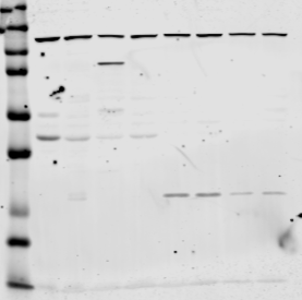
- Bands at high weights
- Bands at low weights
- Uneven or distorted bands
Wrong band size

Protein Variants |
Use the proper control to detect the correct band size. A recombinant protein or overexpression lysate, and/or a peptide competition of the primary antibody may be used. |
No Bands/Signal

Antibody concentration |
Increase the concentration of the primary or secondary antibody. |
Non-specific binding of primary antibody |
Use the blocking step just prior to primary antibody incubation. |
Protein concentration |
Titer the total protein lysates until the signal appears. It is best to use a positive control lysate to show expression in the the target protein, or an over-expression lysate, or a recombinant protein. Use a fraction specific prep to increase the concentration of the protein such as a nuclear prep vs. a whole cell prep. Make sure that the lysis buffer used was strong enough to disrupt the cell's membrane. Ensure complete protein transfer to the membrane by Ponceau staining and that the proteins have left the gel by Coomassie staining. |
Antibody compatibility |
Ensure that the primary antibody reacts with the species of the lysate and it is suitable for western blot. Make sure the primary antibody reacts with the secondary antibody. |
Blocking |
Lower the concentration of the blocking agent or lessen the incubation time. |
ECL Reaction |
Always use fresh reagents. Test by dot blotting the secondary antibody onto membrane and incubating with ECL. |
Image exposure |
Increase exposure time of the film. |
Native proteins |
Some antibodies can not be used on reduced or denatured proteins. Run blot with proteins in native conformation, without SDS. |
Small proteins |
Use smaller pore size membrane (0.2um instead of 0.45um) to prevent transfer through the membrane. |
Large proteins |
Use lower percent gel and use a lower voltage to transfer overnight at 4C. Add SDS to transfer buffer and lower the concentration of methanol. |
Sodium Azide |
Remove from all buffers as it inhibits the HRP reaction. |
High Uniform Background

Insufficient blocking |
Block for one hour at room temp, use 3 % - 5 % BSA instead of milk. |
Insufficient washing |
Wash for five minutes three to fives times on a high speed orbital shaker. Use a large volume of wash buffer. |
Non-specific binding |
Lower the concentration of primary and secondary antibodies by diluting in blocking solution. Incubate primary antibody overnight at 4C. Confirm secondary is specific by omitting the primary, and running a control blot. |
Incompatible block |
Do not use milk if you are using a phospho-specific antibody or an avidin/strepavidin secondary. Milk contains biotin and casein. Do not use milk or BSA if secondary is anti-bovine, anti-goat, or anti-sheep. This will the secondary to recognize the bovine IgG in the block. Use 5 % serum from host animal of the secondary antibody. |
Dry membrane |
Do not allow membrane to dry out after applying the antibody, keep covered with parafilm. |
Film exposure |
Reduce time or wait fifteen minutes before exposing. |
ECL incubation |
Reduce development time by 1-3 minutes. |
Swirly Speckled Background
Mishandling of membrane |
Try to minimize contact of the membrane and gel. Use clean tools to touch membrane and gel. Do not use hands to directly touch the membrane. |
Buffer contamination |
Use fresh buffers for every experiment. Make sure to filter the buffer before use. |
Uneven plastic wrap |
Use sheet protectors instead of plastic wrap. They are made up of tougher plastic and easier to uniformly flatten. |
Air bubbles |
Gently roll out all air bubbles between the membrane and the gel prior to starting the transfer step. |
HRP aggregation |
Filter the secondary antibody with 0.2um filter to remove aggregates. |
Insufficient washing |
Wash for five minutes three to fives times on a high speed orbital shaker. Also try to use a larger volume of wash buffer. |
White Bands

Too much antibody/protein |
White bands surrounded by black are caused by intense localized signal that completely exhausts the ECL reaction with a quick burst of light. No light is then produced during development and a white band occurs. Use a more diluted primary, secondary, or protein concentration. |
Smeared Bands/Lanes
Protein load |
Use less protein and/or try an immunoprecipitation to enrich your target in the lysate. |
Antibody concentration |
Dilute the antibody concentration. |
Bands at High Weights
Protein may form multimers |
This is likely if you see extra bands at high molecular weights that are 2x or 3x the weight of the expected bands. Some proteins will form dimers, trimers, or larger multimers due to disulfide bond formation if the samples are insufficiently reduced. To prevent this, try boiling the sample for longer in Laemmli buffer during sample preparation. |
Bands at Low Weights

Proteases may have digested the protein |
Add protease inhibitors to prevent protein degradation. |
Uneven or Distorted Bands

Voltage may have been too high during migration |
If the voltage is too high, migration will occur too quickly. Check the protocol for the suggested voltage and decrease if necessary. |


